Transthoracic Echocardiogram (TTE)
This is the standard, most commonly used method of echocardiography. Views of the heart are obtained by moving the transducer to different locations on the chest or abdomen wall.
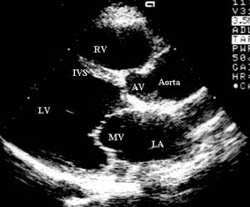
This is a photo of the normal heart seen in the parasternal long axis view.
Right ventricle (RV), inter-ventricular septum (IVS), aortic valve (AV),
Aorta, left ventricle (LV), mitral valve (MV) and left atrium (LA) is seen.
Echocardiography
Test Overview
Echocardiography (sometimes called an echo or an echocardiogram) is a type of ultrasound test that uses high-pitched sound waves to produce an image of the heart. The sound waves are sent through a device called a transducer and are reflected off the various structures of the heart. These echoes are converted into pictures of the heart that can be seen on a video monitor.
An echocardiogram is used to evaluate how well your heart chambers fill with blood and pump it to the rest of the body. Echocardiography can also be used to estimate the amount of blood pumped out of your left ventricle with each heartbeat (called the ejection fraction). An echo also helps evaluate heart size and heart valve function.
Echocardiography can help identify areas of poor blood flow in the heart, areas of heart muscle that are not contracting normally, previous injury to the heart muscle caused by impaired blood flow, or evidence of heart failure, especially in people with chest pain or a possible heart attack. In addition, echocardiography can identify a pericardial effusion and certain congenital heart defects.
Atherosclerosis
Coronary Artery Disease
What is coronary artery disease? Coronary artery disease (CAD) affects the coronary arteries, the blood vessels that supply blood to your heart muscle. These arteries become narrowed or blocked due to fat (cholesterol) and calcium buildup inside of them, which forms a plaque. This process is called hardening of the arteries, or atherosclerosis. Plaque buildup in the coronary arteries reduces blood flow to your heart muscle. Poor blood flow to the heart muscle is called ischemia.
What causes coronary artery disease?
The process of plaque buildup in your coronary arteries, which reduces blood flow to the heart muscle, is called atherosclerosis. Certain risk factors can increase your chances of developing CAD due to atherosclerosis. Smoking, uncontrolled high blood pressure, high cholesterol, diabetes, increased age, male sex, and a family history of heart disease are strong risk factors for coronary artery disease. If you are overweight, respond poorly to stress, and do not exercise regularly, you may also be at an increased risk for coronary artery disease. See the What Increases Your Risk section for more information.
How will I know if I have coronary artery disease?
Chest pain or discomfort is the most common symptom of coronary artery disease (CAD), although it can have many different causes. The chest pain or discomfort associated with CAD is caused by poor blood flow and reduced oxygen delivery to the heart muscle (ischemia) and is called angina. See the Symptoms section for more information.
Not everyone with coronary artery disease has symptoms. A recent study found that as many as a third of people diagnosed with heart attack did not experience chest discomfort.
What can I expect if I have coronary artery disease? You may be able to manage your coronary artery disease (CAD) with medications and lifestyle changes. Sometimes a procedure is needed to help restore normal function of the coronary arteries. Coronary artery bypass surgery is the most invasive type of procedure that may be used. Other common treatment options are angioplasty(which usually includes stenting) or atherectomy. If CAD progresses, you may develop additional problems. Over time, reduced blood flow may weaken your heart muscle so that it is not able to pump effectively. This may result in serious complications, such as heart failure or arrhythmias.
If a plaque breaks open (ruptures), a blood clot may form and suddenly block the blood flow to your heart muscle, causing a heart attack (myocardial infarction). When deprived of blood, oxygen, and nutrients, heart muscle cells die.
Atherosclerosis can affect other arteries throughout your body. Arteries that are commonly affected include those that supply blood to your heart, brain (cerebrovascular disease), and limbs (peripheral vascular disease). See the topics Stroke and Peripheral Vascular Disease of the Legs for more information.
How will a doctor diagnose coronary artery disease?
Your doctor will evaluate your risk factors and symptoms with a medical history and physical exam. If coronary artery disease (CAD) is suspected, you may have additional tests to determine the diagnosis. The most common initial tests to evaluate possible CAD are electrocardiography (EKG or ECG), chest X-ray, and exercise electrocardiography. You may have additional tests if more information is needed or if you are at a high risk for having a heart attack.
What can I do to live with coronary artery disease? If coronary artery disease (CAD) is detected and treated early, it may be reversible to some degree. You can also take measures to prevent a heart attack.
Many studies support lowering average to high cholesterol levels to help reduce your risk of heart attack, stroke, and death. Lowering cholesterol levels appears to benefit you most when you also have heart disease or if you are at increased risk for heart disease.
If you already have coronary artery disease that is causing problems, medication or surgery may help relieve symptoms. These treatments may also prevent a heart attack, the development of heart failure, and early death.
Who can treat my coronary artery disease?
Call your primary care doctor (such as a family practitioner or internal medicine doctor) or a cardiologist if you think you have symptoms of coronary artery disease or if your symptoms worsen. Seek emergency medical care if you have signs of a heart attack, stroke, or sudden heart failure.
Beating Heart Ultrasound
The Test
Echocardiogram An Echo uses sound waves to form a picture of the heart valves and heart muscle. The Echo machine sends sound waves to a transducer (a sound sensitive instrument) that is placed on the chest. The sound waves are reflected by the heart walls (muscle) and heart valves, back to the transducer, which changes the sound into a picture.
Purpose: To determine information about heart chamber size, wall motion, valve movements, and structural changes in and around the heart.
Preparation: There is no special preparation for this test.
Procedure: You will be asked to remove clothing above the waist and put on a gown. EKG patches will be placed on your chest. You will be lying on your back or left side. A technician will apply gel to your chest and a transducer will be placed over the heart area. An EKG will be recording the electrical activity of your heart. A DVD/CD, and a printed report page with ultrasound pictures will be provided for you to take to your physician.
Time: An Echo takes about 15 to 20 minutes.
Beating Heart